http://www.asyura2.com/22/iryo9/msg/346.html
| Tweet |
「EXCLUSIVE: Shocking microscopy photos of blood clots extracted from those who “suddenly died” ? crystalline structures, nanowires, chalky particles and fibrous structures」
(By Mike Adams 2022/6/12)
https://naturalnews.com/2022-06-12-blood-clots-microscopy-suddenly-died.html
独占記事:「『突然死』した人から抽出された血栓の衝撃的な顕微鏡写真
- 結晶構造、ナノワイヤー、チョークのような粒子、繊維状構造物」
(Natural News) 独占記事: 本日、我々は、コビドワクチン接種後、通常数ヶ月で「突然死」した大人から当たり前のように発見されている奇妙な血塊の顕微鏡写真を公開する。
これらの塊はしばしば「血栓」と呼ばれるが、通常の血栓とは全く異なり、単なる血球をはるかに超えるもので構成されている。ゼラチン状でほとんどゼリーのような通常の血栓とは異なり、これらのいわゆる「塊」は、極めて大きく複雑な反復構造要素(すべて以下に示す)を含んでおり、これらの血栓によって死亡した犠牲者の血液の中で明らかに構築されているのである。
これらの血栓はすべて、死後数時間以内に患者から採取されたものである。これらは死後のうっ血によるものではない。これらは血管や動脈に見られる構造物である。凝固した血液ではない。
この血塊を提供してくれた死体防腐処理者(Richard Hirschman)を紹介してくれたJane Ruby博士に、公式に感謝したい。(テレグラムチャンネル T.ME/DRJANERUBY) ルビー博士の執念がなければ、このレポートはなかっただろう。Ruby博士はStew Peters Show (StewPeters.TV)に頻繁に出演しており、月曜日のInfowars.comの放送でも私のゲストとして紹介される予定である。
この生の血栓を、血液を洗って保存し、染色する前の小瓶がこちら。
これらの構造は、以下のような驚くべき特性がある:
・ 丈夫で繊維状、弾力性があり、小さな輪ゴムに似た素材特性を示す。
・ 小さな線維性糸状体が何本も連なったものである。
・ これらの線維束(下の最後の写真参照)は、まるで体が血管の中に別の生命体を作るようプログラムされているかのように、鱗片状の工学的パターンの繰り返しを示している。
・ これらの血栓には奇妙な結晶のような構造があり、透明で通常のグラム染色に耐性を示す。
・ 下の写真は、シリコンに似た生体回路やマイクロチップのように見える構造の一例である。それが何であるかは不明。
・ 下の写真セットの1つには、バイオサーキット・ワイヤーらしきものが写っており、反復パターンとナノスケールの界面構造がはっきりと表れている。これらは目的不明だが、特定の形状に組み立てられている。
これらの写真の背景:
・ この「血栓」のサンプルを、死体防腐処理業界で活躍していて信頼できる死体防腐処理者(Richard Hirschman)から受け取り、これが血管でもその他の組織でもないことを確認した。死体防腐処理の際に血管の内側から取り出された構造物である。
・ 私はこれらのサンプルを、顕微鏡観察時にコントラストを高めるために、微生物学で用いられる標準的なグラム染色法を用いて染色した。下のサンプルのうち、より黄色っぽいサンプルは、ヨウ素のみで染色し、紫色の染色は一切使わなかった。
・ その後、エチルアルコールで洗浄し、標準的な顕微鏡用組織試料作製法を用いてスライドを作製した。
・ 顕微鏡の倍率は20倍から1500倍までで、写真によって異なる。倍率は各写真に示されている。
・ これらのサンプルは私が所有しており、必要であればこれらの写真を再撮影することができる。また、実験室の顕微鏡オペレーターであれば、同じサンプルを使って、これらの写真を再現することができる。
・ 以下に示す説明は私自身の観察に過ぎず、同定された物質の確実性を示すことを意図するものではない。例えば、私が「生体回路」や「ナノワイヤー」について話すとき、これらが実際に生体回路を目的として設計された構造であることを確認することはできない。ただ、そのように見える構造に似ているだけで、それを確認するためにはさらなる研究が必要である。
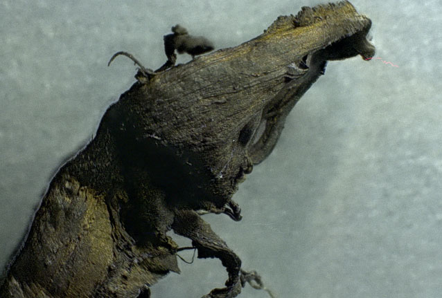
■ 顕微鏡写真セット#1: 不思議な結晶のようなナノ構造
この最初のセットは、奇妙な結晶のような構造を示す。染色できず、通常、血液や血栓には見つからないある種のナノスケールの透明な結晶構造を示しているようだ。
これらの写真で見ているものはすべて、亡くなった人間から採取された血栓の一部である。
倍率は、20倍、50倍、200倍、500倍。
■ 顕微鏡写真セット#2: 構造、より糸、粒子
このセットでは、血栓のより糸、構造、粒子の詳細をクローズアップしている。
倍率は20倍、50倍、100倍、200倍、500倍、1000倍 (極端に拡大すると被写界深度が浅くなるため、高倍率の写真は部分的にぼやけて見える)。
■ 顕微鏡写真セット#3: 結晶状構造体
樹皮状の血栓に結晶のようなものが付着している。この血栓は、紫色の染色剤で染色されているため、濃い紫色をしていることに留意してほしい。
倍率は、20倍、50倍、100倍、200倍、500倍、1000倍。
■ 顕微鏡写真セット#4: 繊維状の物質は、単に血球が凝固したものではない
次のサンプルは、ヨウ素で染色した後、エチルアルコールで洗浄したもの。これがどこから来たのか知らなければ、ビーフジャーキーやチキンナゲットのサンプルだと勘違いするかもしれない。しかし、これはすべて血管や動脈の中にあった血栓の組織である。
ご覧の通り、これらは決して「普通の」血栓ではない。これらは構造を持っており、繊維状である。これは明らかに体内で作られたもので、タンパク質合成の指示によって、ほとんど筋肉組織に似たこの大きな塊を作り出している。繰り返すが、これは血管の中で作られたものである。
倍率は、20倍、50倍、100倍、200倍。
■ 顕微鏡写真セット#5: シリコンに似たチップ構造
このシリーズでは、シリコンベースのマイクロチップ構造に似たものを撮影しているが、これが何かの回路であると断言することはできない。マイクロ回路が同じ倍率でこんなふうに見えるだろうということ。
倍率は20倍、50倍、100倍、200倍、500倍。
■ 顕微鏡写真セット#6: チョーク状の白い粒子
ある死体防腐処理者によると、この人たちの死体から排出された血液には、しばしば「チョークのような」白い粒子が見つかるそうで、肉眼でも見えることもあるという。
私の顕微鏡写真は、このチョークのような白い粒子を捉えている。染色されず、これらの血塊の特定の領域に散らばっているようである。
倍率は、20倍、50倍、100倍、200倍、500倍、1000倍、1500倍。
■ 顕微鏡写真セット#7: 「ナノワイヤー」構造と繰り返される鱗状構造
この写真は、一見、マイクロスケールのワイヤーに見える。拡大してみると、上部にナノスケールのワイヤの界面接合と思われる一連の繰り返し構造が見える。この「ワイヤー」全体は反復するセグメントでできており、その外層は反復する「鱗状」パターンで覆われている。それは、人間ではなく爬虫類の皮膚に似ている。
ちなみに、この構造物が何であるかは分かっていない。しかし、これが循環器系のどこにも属さないことは明らかだ。
最後に、この繊維は単なる人間の髪の毛ではない。血栓にしっかりとくっついていて、取り除こうとしても簡単にはちぎれない。これは異物混入の問題ではなく、血栓そのものから出てきた構造物である。ここにあるものはすべて、人間の血管から出てきたものなのである。
ここで使用した倍率は、20倍、50倍、100倍、200倍、500倍、1000倍、1500倍。
■ いったい、これらは何なのか?
これらの構造が何であるかはまだ分かっていない。しかし、それらが何でないかはわかっている。単なる凝固した血球ではないのである。もしそうであれば、上の最後の写真のように1500倍の倍率で、個々の血球を見ることができるはずである。これは血球ではなく、タンパク質の構造体なのだ。
このように血液中を循環しているタンパク質の構造が、時間をかけて構築されているのは、明らかに体内の細胞によるものだ。細胞内のリボソームが、どのようなタンパク質を構築すべきかを体に指示しているのである。このリボソームが、mRNA遺伝子治療注射によって乗っ取られ、細胞に新しい指示を上書きし、人間以外のものを製造するようになってしまうのだ。
このような構造は、「ワクチン」という偽の傘の下で人々に注入されたmRNAのタンパク質合成指示の結果であると私は考えている。私は、これがどこから来ているのかについて、他の理論や説明ができる専門家からの情報を待っている。
これらの構造の機能と構成を確認するためには、さらに多くの研究が必要である。しかし、現在世界中に存在する極端な検閲と「科学権威主義」のために、どの研究所や大学も、あえてこれらの血栓を検査しその結果を正直に報告することはない。そうすればNIHからの研究資金や政府の助成金を失うからだ。というのは、ワクチンや生物兵器を開発するのと全く同じ人々が、アメリカのほとんどの科学資金を支配しているからである。
したがって、独立した科学者、研究所、ジャーナリストだけが、あえてこの血栓について真実を語ることになる。
結論から言えば、これらは「血」栓ではない。血液の中の構造物である。「構造的な血栓」あるいは「繊維状の血栓」であり、非常に大きく、時間をかけて体内で構築されていくものだ。
私の重大な懸念は、mRNAを注射されたすべての人が、今この瞬間にも体内でこれらの繊維構造物を構築しているかもしれないということであり、それが主要な動脈を塞ぎ、心臓発作や脳卒中、あるいは「成人突然死症候群」(SADS)の急性原因を引き起こすのは時間の問題だろうということだ。
一見、健康そうに見える多くの大人が突然死するのは、この構造物のせいかもしれないと私は考えている。
詳しくはBrighteon.comとInfowars.comをご覧ください。
これらの調査結果については、月曜日の昼間に、Brighteon.comの私のチャンネルで配信しているポッドキャスト「Situation Update」で詳しく説明する予定。
https://www.brighteon.com/channels/hrreport
さらに、6月13日(月)午前11時(中央時間帯)から始まるInfowars.comの「アレックス・ジョーンズ・ショー」放送で、これらの写真と調査結果をすべて発表する。この3時間の番組では、複数の専門家ゲストがこれらの調査結果についてコメントし、これらが何であるか、そして今どれだけの人々が影響を受けているかについて、彼ら自身の情報を提示する予定である。
www.InfoWars.com をご覧ください。そして人類のために祈りましょう。
-------(翻訳ここまで)----------------------------------------------
たくさんの写真がアップされているので、元記事をご覧ください。
これをきっかけに、多くの研究者が、新型コロナワクチンで体内に生じる異物について
取り組むことを希望します。
(関連情報)
「カナダの研究者が、モデルナとファイザーのCovid注射剤にカーボンナノテクとツリウムを発見
(The Expose)」 (拙稿 2022/5/30)
http://www.asyura2.com/22/iryo9/msg/290.html
「遺体の血管から引き出した白い塊は、体外でも成長した (Stew Peters Network)」
(拙稿 2022/5/15)
http://www.asyura2.com/22/iryo9/msg/214.html
「【閲覧注意】ワクチン接種者の静脈と動脈で発見された不可解な血栓と白い繊維質の物体
Richard Hirschman Dr Jane Ruby (マタタビの羅針盤)」 (拙稿 2022/2/23)
http://www.asyura2.com/21/iryo8/msg/610.html
「世界中の科学者が、白い血栓の謎に取り組み中」 (拙稿 2022/2/5)
http://www.asyura2.com/21/iryo8/msg/467.html
「遺体防腐処理者が、動脈・静脈に見たことのないゴム状の塊が詰まっているのを見つける
(Dr. Jane Ruby Show)」 (拙稿 2022/1/27)
http://www.asyura2.com/21/iryo8/msg/390.html
-------(原文引用ここから)-----------------------------
(Natural News) EXCLUSIVE: Today we are publishing a series of lab microscopy photos of bizarre clots which are now being routinely found in adults who “suddenly died,” usually in a number of months following covid vaccinations.
These clots are often referred to as “blood clots” but they are nothing at all like normal clots, and they consist of far more than mere blood cells. Unlike normal clots which are gelatinous, almost jelly-like, these so-called “clots” contain extremely large, complex, repeating structural elements (all shown below) that are clearly being constructed in the blood of the victims who died from these clots.
All of these clots were extracted from patients within a few hours of their death. These are not the result of post-mortem blood stasis. These are structures found in blood vessels and arteries. They are not congealed blood.
We wish to publicly thank Dr. Jane Ruby for connecting us to the embalmer (Richard Hirschman) who provided these clots. (Telegram channel T.ME/DRJANERUBY) Without the persistence of Dr. Ruby, you would not be seeing this report. Dr. Ruby is frequently featured on the Stew Peters Show (StewPeters.TV) and will also be my featured guest Monday on the Infowars.com broadcast.
Here’s a vial of these raw clots, washed of blood and preserved, before staining:
These structures exhibit the following shocking properties:
They are tough, fibrous and resilient, showing material properties similar to small rubber bands.
They consist of many strands of small, fibrous strands.
These fibrous strands (see the very last photo set below) show repeating patterns of scale-like engineering, as if the body has been programmed to build another life form inside the blood vessels.
There are strange crystalline-like structures found on these clots, exhibiting transparency and resistance to normal gram staining techniques.
Below, you will find one example of a structure that appears to resemble a silicon-like biocircuitry or microchip-like structure. We don’t yet know what it is.
One of the photo sets below reveals what appears to be a biocircuitry wire which clearly shows repeating patterns and nano-scale interface structures that are assembled in a specific geometry for an unknown purpose.
Context for the photos you are about to see:
I received these “blood clot” samples from a reputable embalmer (Richard Hirschman) who is active in the field of embalming and who confirmed these are not blood vessels or other tissues of any kind. They are structures that were evacuated from inside blood vessels during embalming procedures.
I stained these samples using standard gram staining techniques used for microbiology in order to enhance structural contrast during microscopy. One of the samples below ― the more yellowish sample ― was stained only with iodine, not any violet-colored stains.
The samples were then washed with ethyl alcohol and prepared on slides using standard tissue sample preparation for microscopy.
Microscope magnification varies from 20x to 1500x, depending on the photo shown below. Magnifications are indicated with each photo set.
I retain possession of these samples and can reproduce these photographs if required. Any competent lab microscopy operator could reproduce these photos using the same samples.
My descriptions shown below are merely my own observations and are not intended to indicate certainty of the substances being identified. For example, when I talk about “biocircuitry” or “nanowires,” I cannot confirm these are structures actually engineered for purposes of biocircuits. Merely, they resemble structures that seem to indicate such a purpose, but further research would be needed to confirm these observations.
Microscopy photo set #1: Strange crystal-like nanostructures
This first set shows strange crystal-like structures that resist staining techniques and appear to show some sort of nano-scale, clear crystalline structures which would normally never appear in blood or blood clots.
Everything you are looking at in these photos is part of a blood clot extracted from an expired human being.
Magnifications shown here are 20x, 50x, 200x and 500x:
Microscopy photo set #2: Structures, strands and particles
This second set shows very close-up details on the strands, structures and particles found in these blood clots.
Magnifications shown are 20x, 50x, 100x, 200x, 500x, 1000x: (extreme magnifications causes a loss of depth of field which is why the highly-magnified photos seem so blurry in certain areas)
Microscopy photo set #3: Crystal-shaped structures
Crystal-like structures are attached to the bark-like structure of the blood clot. Remember, this clot is stained using a violet stain, which accounts for its dark purple color.
Magnifications are 20x, 50x, 100x, 200x, 500x and 1000x:
Microscopy photo set #4: Fibrous material is not simply congealed blood cells
The following sample was stained with iodine, then washed with ethyl alcohol. If you did not realize where this came from, you might think this was a sample of beef jerky or a chicken nugget. In reality, all of this is clot tissue that was found inside blood vessels or arteries.
As you can see, these are in no way “normal” blood clots. These have structure and are fibrous. They are clearly being built by the body, using protein synthesis instructions to create this large mass that nearly resembles muscle tissue. Yet it is being built inside the blood vessels.
Magnifications are 20x, 50x, 100x and 200x:
Microscopy photo set #5: Silicon-like “chip” structure
This series shows something that appears to resemble silicon-based microchip structures, although I cannot claim with certainty that this is a circuit of any kind. It simply resembles what micro-circuitry looks like at similar magnifications.
Magnifications used here at 20x, 50x, 100x, 200x and 500x:
Microscopy photo set#6: Chalk-like white particles
An embalmer told me that blood emptied from the bodies of these people during embalming often appears to show “chalk-like” white particles which are visible even to the naked eye in certain cases.
My microscopy photos seem to have captured some of these chalk-like white particles which resist staining and seem to be scattered across certain regions of these clots.
Magnifications used here are 20x, 50x, 100x, 200x, 500x, 1000x and 1500x:
Microscopy photo set#7: “Nanowire” structures and repeating, structural scales
What follows here is a stunning look at what appears to be, at first, a micro-scale wire. Zooming it, we see a series of repeating structures along the top that appear to be nano-scale wire interface junctions. The entire “wire” is made of repeating segments, and its outer layer is covered in repeating “scale-like” patterns that actually resemble reptile skin more than anything human.
For the record, we don’t know what these structures are. However, it’s clear this doesn’t belong anywhere in the circulatory system.
Finally, this fiber is not simply a human hair. It is firmly attached to the blood clot and when I tried to remove it, it would not tear away easily. This is not a contamination issue, it is a structure emanating from the clot itself. Everything you see here came out of a human being’s blood vessels:
Magnifications used here are 20x, 50x, 100x, 200x, 200x, 500x, 500x, 500x, 1000x and 1500x:
What is all this?
We don’t yet know what all these structures are. We know what they are not, however: They are not simply clotted blood cells. If they were, then at the 1500x magnification shown in the last photo, above, we would be able to see individual blood cells. These are not blood cells, they are protein structures.
Protein structures circulating in the blood like this, building up over time, are clearly being constructed by the body’s cells. The ribosomes in the cells instruct the body what proteins to construct. These ribosomes are hijacked by mRNA gene therapy injections, which overwrite new instructions to the cells, causing them to manufacture something other than human.
I believe the structures you are seeing above are the result of mRNA protein synthesis instructions which have been injected into people under the false umbrella of “vaccines.” I welcome input from other experts who may have other theories or explanations of where this is coming from.
More research is needed to confirm the function and composition of these structures, yet because of the extreme censorship and “science authoritarianism” that now exists in the world, no lab or university will dare examine these clots and honestly report the results. To do so would risk losing all NIH funding and federal grants, since the very same people who engineer vaccines and bioweapons also control most science funding in America.
Thus, only independent scientists, labs and journalists will dare tell the truth about these clots.
In conclusion, they are not “blood” clots. They are structures in the blood. They are “structural clots” or “fibrous clots” that are extremely large and are being constructed inside the body over time.
My grave concern is that every person who has been injected with mRNA instructions may be constructing these fibrous structures inside their bodies at this very minute, and that it’s only a matter of time before they block major arteries or cause heart attacks, strokes or other acute causes of “Sudden Adult Death Syndrome” (SADS).
I believe these structures may very well explain why so many seemingly healthy adults are suddenly dying.
Hear more details on Brighteon.com and Infowars.com
I will be discussing these findings in more detail Monday mid-day on my Situation Update podcast at my channel on Brighteon.com:
https://www.brighteon.com/channels/hrreport
In addition, I am unveiling all these photos and findings as I host the Infowars.com “Alex Jones Show” broadcast Monday, June 13th, beginning at 11 am central. The three-hour show will feature several expert guests who will comment on these findings and present their own information about what these may be and how many people are being affected right now.
Tune in at www.InfoWars.com
-------(原文引用ここまで)-----------------------------
|
|
|
|
最新投稿・コメント全文リスト コメント投稿はメルマガで即時配信 スレ建て依頼スレ
|
|
 スパムメールの中から見つけ出すためにメールのタイトルには必ず「阿修羅さんへ」と記述してください。
すべてのページの引用、転載、リンクを許可します。確認メールは不要です。引用元リンクを表示してください。